What can larval morphology teach us about ant evolution and development?
In their recently published paper, Di Li and her co-authors illuminate the internal and external morphology of Ooceraea biroi, an important model organism in myrmecological research. Here, the first author tells us about her transition from Neuroptera to Hymenoptera, talks about the joys and difficulties of international collaboration, and shares the story behind the “rainbow sausage” image.
Interview compiled by Salvatore Brunetti and edited by Alice Laciny.

MNB: Could you tell us a bit about yourself?
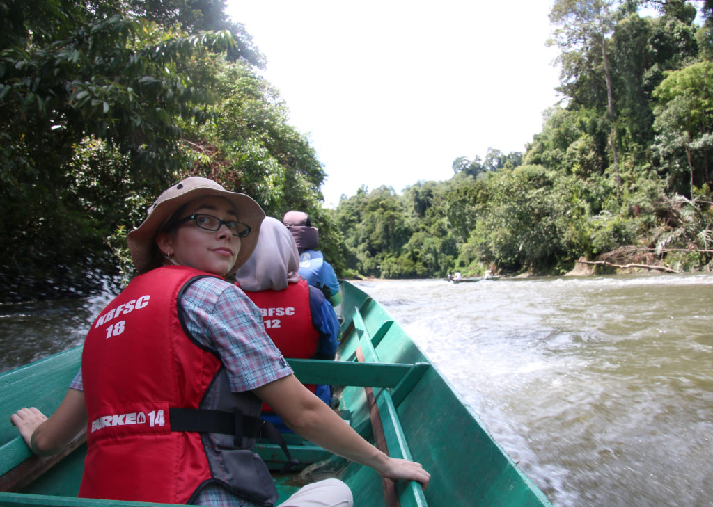
DL: I’m a researcher from China with a background in Neuroptera, particularly the lacewing families Berothidae and Dilaridae. Just a few years ago, I never would have imagined submitting a paper to Myrmecological News! During my PhD, I focused on the taxonomy and phylogeny of these groups. In 2020, I had the chance to spend over a year as a visiting PhD student at the Jena University in Germany. There, I was introduced to 3D reconstruction techniques for anatomical studies, and more importantly, I met many wonderful people—including Rolf, Brendon, and Adrian—who are now co-authors on this paper. Thanks to a series of lucky encounters, they welcomed me into their ant research group, which marked the beginning of my journey into studying Hymenoptera. After completing my PhD, I’ve continued to collaborate with this amazing group on projects involving ants and other Hymenoptera.

MNB: Could you briefly outline your research on “The larval morphology of the clonal raider ant, Ooceraea biroi (Forel, 1907) (Dorylinae), with broader implications for the Formicidae and Hymenoptera” in layman’s terms?
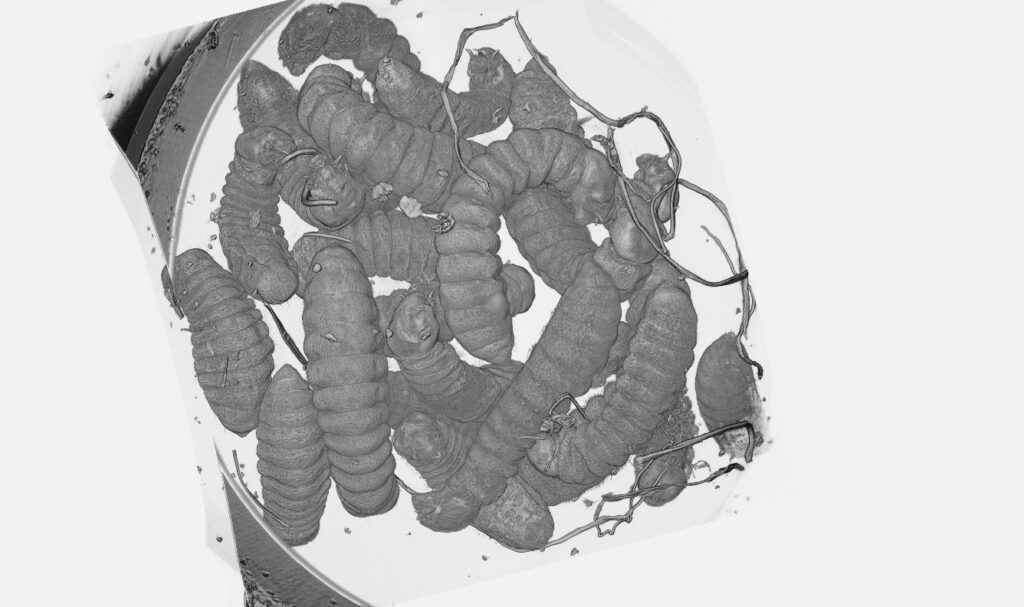
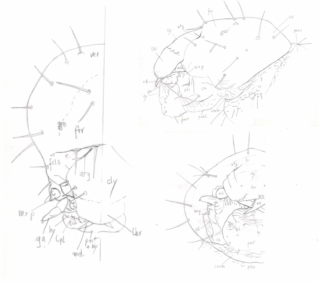
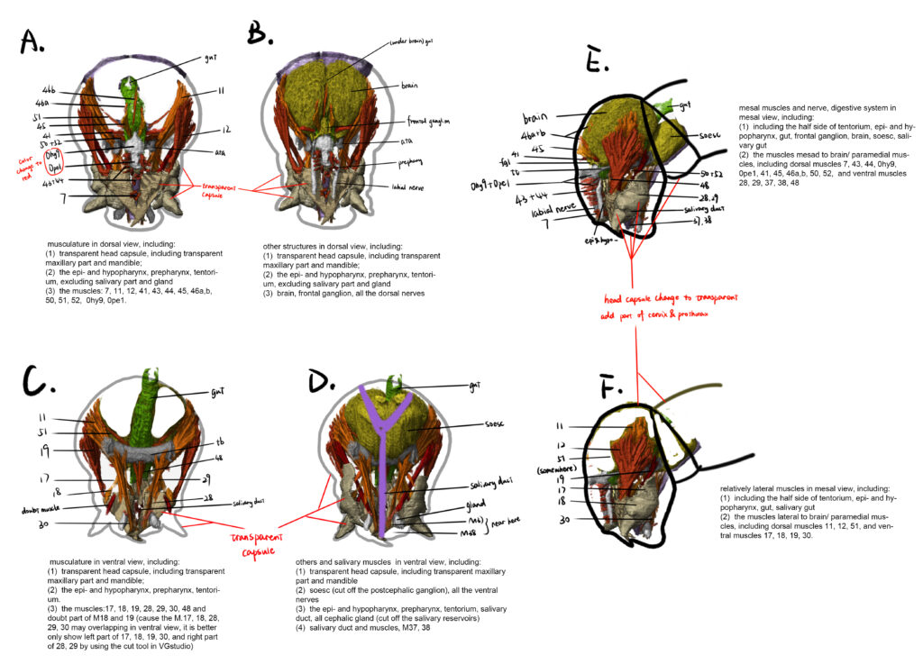
DL: In this study on ant larvae, we provide the first detailed description of both the internal and external morphology of the model species Ooceraea biroi. The larva of this species is grub-like and depigmented, with strongly reduced head structures and a weakly sclerotized, highly simplified, sausage-shaped body. We also compare the documented larval features with those of other Formicidae, broader Hymenoptera, and even larvae from other holometabolous insect orders. Finally, we summarize known morphological characters of ant larvae from previous studies, highlighting the often underestimated diversity and evolutionary significance of larval morphology.
MNB: What is the take-home message of your work?
DL: Our study underscores the importance of larval morphology for understanding ant and Hymenoptera evolution. Phenomic data—large-scale morphological data, i.e., CT scans—hint at the significance of larval form not only as a developmental stage but also as a source of key morphological characters. Although larval forms are often regarded as relatively conserved, our work shows that the complexity of larval morphology, hence diversity, has been underestimated. Exploring this overlooked diversity could help us gain novel insights to understand evolutionary patterns across Hymenoptera.

MNB: What was your motivation for this study?
DL: Honestly, my initial motivation was quite simple—I wanted to understand the complete internal anatomy of holometabolous insect larvae. The story of this paper began shortly before I left Jena more than two years ago. Brendon had proposed documenting the full morphology of Ooceraea larvae, noting that despite advances in morphological techniques, the internal anatomy of Hymenoptera larvae remained largely unexplored. I still vividly remember that moment: while working on a paper about neuropteran larvae, I overheard Brendon and Rolf discussing the Ooceraea larva project nearby. At the time, although I was already familiar with larval cephalic anatomy, I had little experience with postcephalic structures. I saw this as a valuable opportunity to learn about the whole-body anatomy under Rolf’s guidance before leaving Jena—so I volunteered to join the project. Initially, my goal was simply to gain experience with postcephalic anatomy. Motivated by the time pressure, I managed to complete the 3D reconstruction in just one week—a pace that still surprises me when I look back! (laughs) Later, when Brendon formally established the “Jena ant group,” I became a member. My motivations gradually shifted and I realized the broader value of foundational morphological work and its importance for future evolutionary and developmental studies, which also had a lasting impact on me and laid the foundation for my current studies on Hymenoptera.
MNB: What was the biggest obstacle you had to overcome in this project?
The biggest challenge for me came after completing the morphological documentation—writing the discussion section. As I had only focused on Neuroptera in the previous several years, moving into Hymenoptera research meant stepping into a vastly larger and more complex field. Comparative analysis required an extensive knowledge base across many groups, and learning a new group in a short time was daunting. My brain was constantly overheating when drafting the discussion: reading massive numbers of papers daily, taking notes, comparing traits across groups, memorizing new taxa names. At the same time, the “Jena ant team” was scattered across the globe: I had returned to China to finish my PhD thesis, Brendon finished his postdoc in Germany and moved back to the USA, Adrian started a postdoc in Japan, and Rolf remained in Germany. Thankfully, with the collaborative efforts of all co-authors, we refined the discussion section and merged everyone’s viewpoints into the final manuscript. I am truly grateful for my amazing and supportive ant group!
MNB: Do you have any tips for others who are interested in doing related research?
DL: If you’re passionate about morphology, I highly encourage you to explore anatomical approaches—especially using modern techniques like micro-CT or Laser Scanning Confocal Microscope (CLSM). There’s still so much hidden information to uncover, particularly in developmental stages that are often overlooked, such as larvae. Larval morphology is not only fascinating in its own right, but also holds key insights into evolutionary and developmental processes. With the help of new imaging tools, we now have the opportunity to revisit these “forgotten” life stages and come up with some exciting new questions.
MNB: Where do you see the future for this particular field of ant research?
DL: Our corresponding author, Brendon, has already accomplished remarkable work on ant morphology. I believe that continued integrative studies combining internal anatomy, external morphology, and evolutionary analysis will greatly advance our understanding of ant development and diversification.


Recent Comments