Best Paper Award 2023
In 2023, Myrmecological News awards for the sixth time the Best Paper of the previous year. From January 9 to January 23, the editorial board and the community could vote for their most favourite papers published in 2023.
It is now our great pleasure to announce the winner of the certificate of this Best Paper 2023 and the voucher worth the Article Processing Charge (700 EUR) of a future contribution in Myrmecological News:
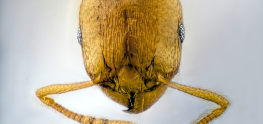
“Wonderfully weird: the head anatomy of the armadillo ant, Tatuidris tatusia (Hymenoptera: Formicidae: Agroecomyrmecinae), with evolutionary implications” written by Adrian Richter et al.
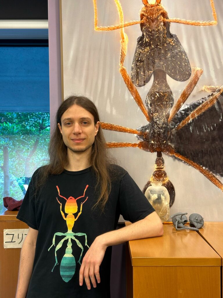
A View by Adrian Richter
I am an insect morphologist from Germany and the current stage of my academic adventure is a postdoc in Okinawa, Japan with Evan Economo at OIST.
My fascination with ants began in my early childhood, so I am thankful and humbled that I am getting a chance to actually study them myself now. My journey in academia began in Jena, Germany, where I did a Bachelor’s, Master’s and PhD with Rolf Beutel.
If I had to summarize my aspirations as a researcher in three words, it would be to “understand ant bodies”. I am using techniques such as micro-CT scanning, electron microscopy and histology to look at the innermost and smallest details of ant anatomy. By comparing different ant species, I want to contribute to understanding how the immense diversity of ants we see on earth has evolved. Recently, I have been trying to incorporate more and more phylogenetic comparative methods in my research, which is mostly focused on the feeding system of ants at the moment. Apart from ants, one thing I really enjoy is Japanese culture and it has been great to visit some of the ancient castles, temples and mountain forests during my time here.
Tatuidris morphology
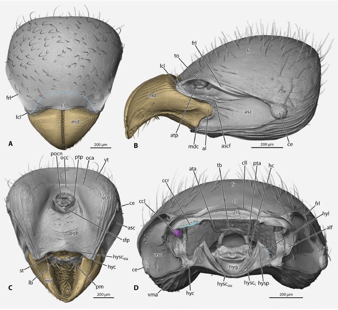
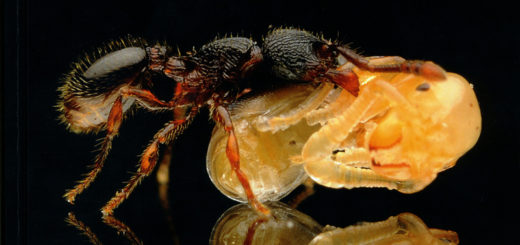
Our paper on the head of Tatuidris tatusia is part of a series of studies I did on ant head anatomy during my PhD project. It was especially fun to reconstruct the head anatomy of this South and Central American species from micro-CT data because it was in many ways very different from all the other ants that I had looked at before. Some of those “weird” features of Tatuidris are their cup-shaped mandibles with a dense brush of thick, spine-like setae and their deep antennal scrobes with antennal sockets that are turned upside down. This unique head shape is also related to some unusual internal features, like an almost right angle in the tentorium, an internal skeletal element that the antennal muscles sit on, which have to reach the upturned antennal socket. We also focused quite a bit on the mandibular articulation because this was, again, rather different from most other ants. Based on 3D-prints of the head and mandibles, we think that there are several stabilizing sliding surfaces that guide the mandible to open along two different axes. The 3D-prints we used to infer this are shown in Figure 11 of the article. Finally, we looked at some of the morphological characters that have been used to describe the different clades of poneroid ants and discussed some of those in the light of the most recent phylogenetic hypotheses. On the one hand, our study is a step on the way to better understand morphological evolution of poneroids and ants in general, and on the other hand, provides some new insights on an ant species so rarely studied, we don’t even know what it eats yet.

That this study got enough appreciation in the myrmecological community to win the best paper award is incredibly humbling, and we are very thankful that our work has found an interested readership.

Indeed, one of the best articles last year. Hoera!